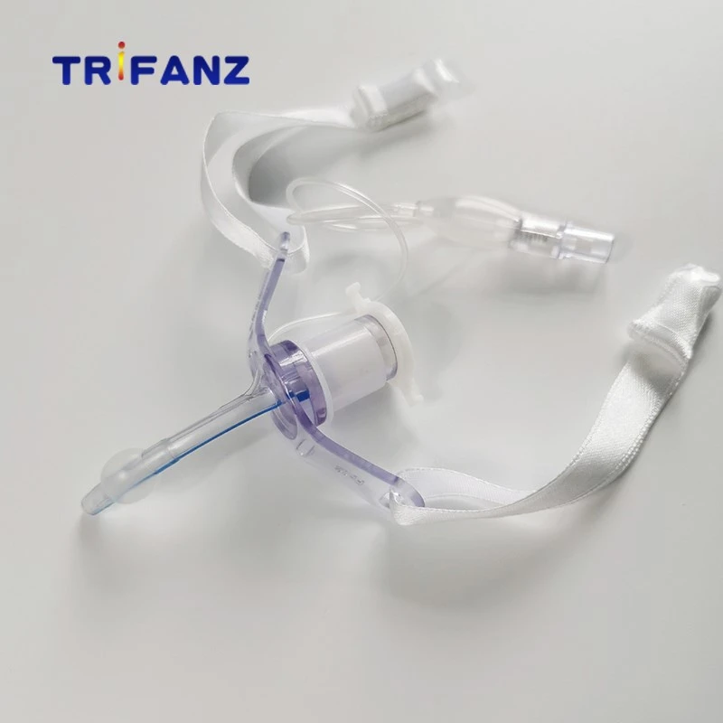


Tracheal intubation and tracheotomy Endotracheal intubationEndotracheal intubation (endotracheal intubation) is an effective measure to relieve upper airway obstruction, to ensure the airway is unobstructed, and to perform artificial respiration. It is already a very important method for clinical rescue of critically ill breathing difficulties.Advantages: ①The equipment is simple, the operation is convenient and the effect is quick and effective. ②It can maintain unobstructed breathing and facilitate the suction of lower respiratory secretions. ③Facilitate oxygen supply, increase the partial pressure of arterial oxygen and discharge excessive carbon dioxide. ④ It is convenient to perform pressurized artificial respiration or mouth-to-tube artificial respiration to increase effective alveolar ventilation.
【Indications】
1. People who need urgent relief of throat obstruction, such as neonatal dyspnea, infant respiratory distress syndrome, acute infectious throat obstruction, acute throat edema, neck lumps or infection swelling that compress the larynx and trachea and cause breathing difficulties.
2. Retention of lower respiratory tract secretions requires timely suction.
3. Respiratory failure caused by various causes requires artificial respiration.
4. In pediatric bronchography and pediatric tracheotomy, tracheal intubation is required first.The tracheal intubation equipment is simple, with anesthesia laryngoscope and intubation (Figure 3-13-1). At present, the clinical application of tracheal intubation includes rubber intubation, polyvinyl chloride intubation and silicone polyethylene intubation. Among them, the silicone tube has the least irritation and the rubber tube has the most irritation.Intubation specifications are divided into 14 numbers, from F (legal system) 10, 12, 14, 16, 18 to F36. Choose different specifications according to different ages. Generally: F10-12 for newborns, F14-16 for infants from 1 to 11 months old, F16-20 for 1-2 years old, F20-22 for 3-4 years old, F22-24 for 5-6 years old, 7~ Use F24 to 26 for 9 years old, F26 to 28 for 10 to 14 years old, F30 to 34 for young and adult women, and F34 to 36 for adult men.
[Intubation approach]
1. Transnasal endotracheal intubationThe advantages include: ①The intubation is not too thick and the chance of damaging the larynx is small. ② Observe the nasal mucosa to understand the reaction to intubation. ③ Better fixed. ④The patient cannot bite the intubation tube and does not hinder swallowing. ⑤Those with difficulty in opening the mouth must be intubated through the nose.Disadvantages include: ① The operation is time-consuming and difficult to succeed. ②The tube length and inner cavity are small, and the dead space is large. It is easy to be blocked by secretions and increase the respiratory resistance. ③It is easy to bring the infection of the nasal cavity into the lower respiratory tract.
2. Oral endotracheal intubationThe advantages include: ① Simple and convenient operation. ②Do not damage the nasal cavity. ③Easy to suck lower respiratory secretions. ④ It is easier to change the intubation.Disadvantages include: ①The intubation tube is not easy to fix, and the sliding of the tube can easily cause throat damage. ②The patient feels very uncomfortable and hinders chewing and swallowing.
[Intubation method]
1. Anesthesia: Children do not need anesthesia. Adults use 1% Decaine to spray the pharynx and larynx as a topical anesthesia.
2. Position: Take the supine position more, with the head slightly raised and back.
3. Method:(1) Oral intubation: Place gauze on the patient's upper incisor. The surgeon's left hand holds the anesthesia laryngoscope or directly extends the laryngoscope to the throat, and sees the epiglottis, the epiglottis is raised, exposing the glottis, and the right hand holds the tip of the intubation tube with a metal guide core (usually a thicker steel wire) in the sound On the door, when the inhalation glottis is opened, the intubation tube is immediately inserted, and the exhalation of gas at the rear end of the tube means that the tube has been inserted into the trachea. After adjusting the intubation tube to an appropriate depth, pull out the metal guide core. Fix the bite stopper and the intubation tube together on the cheek.(2) Nasal intubation: Choose an appropriate type of nasal intubation, apply lubricant to the outside of the tube, enter the tube through the nasal cavity, through the nasopharynx and oropharynx, adjust the position of the head, and insert the tube into the trachea through the throat. When intubation is difficult, an anesthesia laryngoscope can be used to insert the intubation through the glottis as described above.(3) Endoscopically guided tracheal intubation: Due to difficulties in opening the mouth, small jaw deformity, etc., it is difficult to expose the glottis under anesthesia laryngoscope, or the oral or nasal intubation fails, this method can be used. Method: After surface anesthesia (1% decaine) of the oropharynx, larynx, and nasal mucosa, use a fiber laryngoscope or fiber bronchoscope to pass through the intubation, and insert the fiber endoscope into the larynx or trachea through the mouth or nose, and then homeopathically The anesthesia cannula is pushed into the trachea under the guidance of the fiber endoscope.When artificial respiration is performed after intubation, it should be observed whether the thoracic expansion on both sides is symmetrical and whether the breath sounds of the lungs on both sides are equal.
[Complications]
Complications of tracheal intubation include abrasions of the larynx and trachea, ulcers, edema, granulation formation, dislocation of prickle cartilage, cylindroid arthritis, and membranous tracheitis. Severe cases can cause throat stenosis, and the causes of complications are: ①The operator is unskilled or careless in operation. ②The quality of the intubation is not good. ③Improper selection of tubes or excessively thick tubes. ④Secondary infection. ⑤The intubation time is too long,
【Precautions】
1. The selected cannula should be small in irritation, suitable in size and well fixed.
2. Aseptic operation to avoid infection.
3. The operation is light and accurate.
4. Do not insert too shallow or too deep, children should enter 2.5~3cm below the glottis, and adults should use 4~5cm.
5. The intubation time should not exceed 72 hours for children and 48 hours for adults. If the blood oxygen does not improve after oxygen and artificial respiration within this time, tracheotomy should be performed.
6. Children should not use cuffed intubation. Adult cuffs should not be over-inflated and deflated for 5-10 minutes every hour to prevent local compressive necrosis.
7. Give adequate fluids and give antibiotics to prevent infection.When using an artificial ventilator after intubation, you should always pay attention to the adjustment of the pressure or volume of the ventilator. When there is no artificial respirator, it is easiest to perform artificial respiration with a compressed air bag. For artificial respiration with pressurized oxygen, the pressure of children should not exceed 30cmH2O. Speed 40 times/min. Each air volume is 20ml. The time ratio between inflating (inhalation) and expiration (exhalation) should be 1:2. If possible, blood gas analysis should be done to understand the effect of artificial respiration. TracheotomyTracheotomy is an emergency operation to rescue critically ill patients. It is an operation to cut the anterior wall of the neck trachea so that the patient can breathe through the newly established passage. It is mainly used to rescue patients with blocked larynx.
[Applied Anatomy]
The cervical trachea is located in the middle of the neck, with skin, fascia, sternohyoid muscle and sternothyroid muscle covering the front. The medial edges of the banded muscles on both sides connect with each other at the midline of the neck to form a white line. When performing tracheotomy, follow this line to separate to the deep part, which makes it easier to expose the trachea. There are about 7 to 8 tracheal rings in the cervical trachea. The thyroid isthmus is generally located in the 2nd to 4th tracheal rings. The tracheal incision should be made at the lower edge of the isthmus to avoid damage to the thyroid and cause bleeding. The innominate artery and vein are located on the front wall of the 7th to 8th tracheal ring, so the incision should not be too low. There is no cartilage on the posterior wall of the trachea, and it is connected with the front wall of the esophagus. When the trachea is cut, it should not be cut too deep to avoid damage to the esophageal wall.The common carotid artery and internal jugular vein are located in the deep part of the sternocleidomastoid muscles on both sides. At the level of the cricoid cartilage, the above-mentioned blood vessels are far from the midline position, and gradually move downward to the midline. They are close to the trachea at the suprasternal fossa. The triangular area on the top and the front edge of the sternocleidomastoid muscle is called the safety triangle. The tracheotomy is performed along the midline in this triangle to avoid damage to the large blood vessels in the neck.