





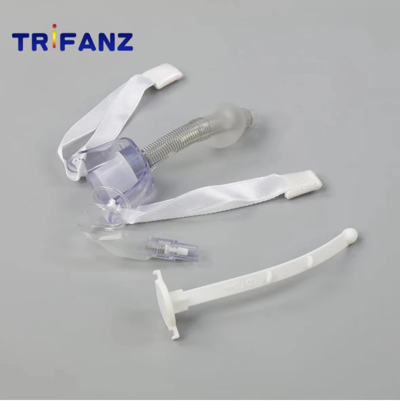


【Indications】
1. Throat obstruction: Throat obstruction of degree III to IV caused by any reason, especially when the cause cannot be resolved quickly.
2. Lower respiratory tract secretions retention: coma, brain disease, nerve palsy, severe brain, chest, abdominal trauma, and respiratory tract burns caused by lower respiratory tract secretions retention. In order to aspirate sputum, tracheotomy can also be performed.
3. Preventive tracheotomy: In some oral, maxillofacial, pharynx, and larynx operations, in order to keep the postoperative airway unobstructed, a tracheotomy can be performed in advance.
4. When assisting breathing for a long time: tracheotomy also provides convenience for the installation of assisted breathing apparatus.
【Preoperative preparation】
1. Prepare surgical instruments including scalpel, scissors, tracheotomy retractor, vascular forceps, forceps, suction device, etc.2. Prepare tracheal tubes according to age and sex. Adult men generally use 10mm pipe diameter, and adult women use 9mm pipe diameter casing.
[Anesthesia]
Local anesthesia is generally used. 1% procaine or 1% lidocaine was injected into the anterior midline of the neck subcutaneously and subfascially.
【Surgical methods】
1. Position: The most suitable position for tracheotomy is supine position, with a pillow under the shoulders, and the head tilted back so that the trachea is raised and close to the skin, which is convenient for exposing the trachea during the operation. But reclining should not be excessive, so as not to aggravate breathing difficulties. If the breathing difficulty is severe and the patient cannot lie supine, the operation can be performed in a semi-recumbent or sitting position, but exposing the trachea is more difficult than in the supine position (Figure 3-13-2).
2. Disinfection: Disinfect the neck skin according to the surgical method. When the condition is very critical, an emergency tracheotomy can be performed immediately without disinfection.
3. Surgical steps:(1) Incision: A straight incision can be used, starting from the lower edge of the thyroid cartilage to near the upper sternal fossa, and incise the skin and subcutaneous tissue along the anterior midline of the neck to the upper sternal fossa (Figure 3-13-3). Or take a transverse incision at the 3CM lower edge of the cricoid cartilage (Figure 3-13-4). (2) Separate the anterior cervical muscle layer: Use hemostatic forceps to make a blunt separation along the midline of the neck. Use a hook to pull the sternohyoid muscle and sternothyroid muscle to both sides with equal force. To maintain the midline position of the trachea, and often touch the cricoid cartilage and trachea with your fingers, so that the operation is always performed along the anterior midline of the trachea (Figure 3-13-5). (3) Exposure of the trachea: The thyroid isthmus covers the front wall of the trachea of the 2nd to 4th rings. If the isthmus is not wide, it is slightly separated at the lower edge and pulled upward to expose the trachea; if the isthmus is too wide, the trachea can be exposed. It is cut and sutured to stop bleeding in order to expose the trachea.(4) Confirm the trachea: After the thyroid is separated, the tracheal ring can be vaguely seen through the anterior tracheal fascia, and the circular cartilage structure can be felt with the fingers. It can be punctured with a syringe, depending on whether the gas is withdrawn, so as not to mistake the large blood vessels in the neck for the trachea in an emergency. If necessary, the cricoid cartilage can be found first, and then dissected down to find and confirm the trachea.(5) Cut the trachea: After confirming the trachea, inject 2ml of 0.5% decaine or 1% lidocaine into the trachea. At the 2nd to 4th rings, use a blade to pick up the 2 tracheal rings from bottom to top (Figure 3-13-6). Or ∩-shaped incision of the anterior wall of the trachea to form a tongue-shaped anterior wall flap of the trachea. The flap is sutured with the subcutaneous tissue and fixed with a needle to prevent the tracheal incision from being difficult to find after the tracheal tube is pulled out or when changing the tube, which may cause suffocation.(6) Insert the tracheal cannula: Use a tracheal dilator or curved hemostatic forceps to open the tracheal incision, insert the selected cannula with a tube core, immediately remove the tube core, and put it into the inner tube (Figure 3-13-7) . If any secretions are coughed up from the mouth of the tube, confirm that the cannula has indeed been inserted into the trachea. If no secretions are coughed up, a little gauze fiber can be placed in the mouth of the tube and see if it floats with the breath. If it is found that the cannula is not in the trachea, the cannula should be pulled out, inserted into the tube core, and reinserted.(7) Fixed sleeve: The two outer edges of the sleeve plate are firmly tied to the neck with cloth tape to prevent it from falling out; the elasticity of the tie should be moderate.(8) Suture: If the neck soft tissue incision is too long, 1-2 stitches can be sutured at the upper end of the incision, but it is not advisable to suture too tightly to avoid aggravating postoperative subcutaneous emphysema.
【Postoperative care】
1. Keep the cannula unobstructed; after the trachea is cut, the sleeve must be kept unobstructed at all times. If any secretions are coughed up, they should be wiped off immediately with gauze. The inner tube should be taken out regularly for cleaning and disinfection. Then reinsert it in time to prevent the secretions from drying out and blocking the outer tube. Generally, the inner casing is cleaned once every 4-6h. If there are too many secretions, increase the number of washings.
2. Keep the lower respiratory tract unobstructed; keep proper temperature and humidity indoors, treat with steam inhalation, or regularly instill a little saline, 0.05% chymotrypsin solution, 1% potassium iodide or antibiotic solution through the tracheal tube to dilute the sputum Liquid, easy to cough up. When necessary, a suction device can be used to suck out the lower respiratory tract sputum.
3. Prevent wound infection: due to sputum contamination, postoperative wounds are prone to infection, so the dressing should be changed once a day. Disinfect the skin around the incision, and if necessary, apply antibiotics as appropriate to control the infection.
4. Prevent the casing from falling out: Too short a casing or too loose a belt to fix the casing can cause the outer tube to fall out. Check whether the casing is in the trachea frequently. If the casing is found to come off, it should be reinserted immediately to avoid suffocation. Within 1 week after surgery, it is not advisable to change the outer tube, so as to avoid accidents caused by the difficulty of intubation because the pretracheal tissue has not yet formed a sinus. If it is necessary to exchange, prepare hooks, vascular clamps and other instruments.
5. Extubation: If the symptoms of blocked larynx and lower respiratory tract secretions have been eliminated, extubation may be considered. Before extubation, the tube should be blocked continuously for 24 to 48 hours. If the patient breathes steadily during activity and sleep, the cannula can be removed, the wound does not need to be sutured, and the wound edge is drawn up with butterfly tape, and it can heal itself after a few days. Observe closely within 1 to 2 days after extubation, and deal with it in time if breathing difficulties occur.
【complication】
1. Subcutaneous emphysema: It is the most common postoperative complication. The main causes of subcutaneous emphysema are: ①When the trachea is exposed, the surrounding soft tissue is stripped too much, ②The tracheal incision is too long, or the pretracheal fascia incision is smaller than the tracheal incision. Air is easy to leak from both ends of the incision; ③After cutting the trachea or inserting the cannula, a severe cough occurs, which promotes the formation of emphysema; ④Suturing the skin incision is too tight. It mostly occurs in the neck, and sometimes extends to the head, chest and abdomen. Most of the subcutaneous emphysema can be absorbed by itself within a few days without special treatment.
2. Pneumothorax: When exposing the trachea, it separates too much downwards and damages the pleura, which can cause pneumothorax. In some cases, due to severe throat obstruction and high negative pressure in the chest, the alveoli rupture during severe coughing, forming spontaneous pneumothorax. Mild pneumothorax can generally be absorbed by itself. If the pneumothorax is obvious and causes dyspnoea, thoracentesis or closed drainage should be performed to expel the gas.
3. Wound bleeding: a small amount of bleeding after surgery, iodoform gauze can be filled around the tracheal cannula, compression to stop bleeding, or add hemostatic drugs as appropriate. If there is more bleeding, check the wound and ligate the bleeding point with sufficient preparation.
4. Difficulty in extubation: The main reasons are: ①If the trachea is cut too high, it will damage the cricoid cartilage and cause throat stenosis; ②The granulation hyperplasia at the tracheal incision or excessive resection of the tracheal cartilage ring will cause tracheal stenosis; ③The primary disease is not cured , Extubation is easy to cause breathing difficulties; ④The tracheal cannula model is too large, and the breathing is not smooth during the blockage test. It should be handled as appropriate according to different reasons.Section 3 cricothyrotomyFor critically ill patients with throat obstruction in need of emergency rescue, cricothyroidotomy can be performed first when the tracheotomy is too late. After the dyspnea is relieved, the conventional tracheotomy can be performed.
[Surgery Points]
First, determine the position of the thyroid cartilage and cricoid cartilage. Make a 3~4cm transverse skin incision between the thyroid cartilage and cricoid cartilage, separate the anterior cervical muscle, make a 1cm transverse incision at the cricothyroid membrane, open the wound with a knife handle or vascular forceps to allow air to enter, and then insert Rubber tube or plastic tube and fix it.
【Precautions】
1. Avoid cutting the cricoid cartilage during surgery to avoid throat stenosis after surgery.
2. The intubation time after cricothyrotomy should not exceed 24 hours, and metal cannulaes should be avoided to prevent the cricoid cartilage from abrading and causing throat stenosis.
3. When the situation is very urgent, use a thick injection needle to pierce the subglottic area directly through the cricothyroid membrane, which can temporarily relieve the symptoms of laryngo obstruction. The puncture depth should be controlled appropriately to prevent the needle from not entering the subglottic area or penetrating the posterior wall of the trachea. If a cricothyroid trocar is available, the trocar can quickly relieve breathing difficulties.
Welcome to ask me at any time.
Please add my WhatsApp/WeChat:+86 19518555422.
My Email: sale4@trifanz.com.